OVERVIEW
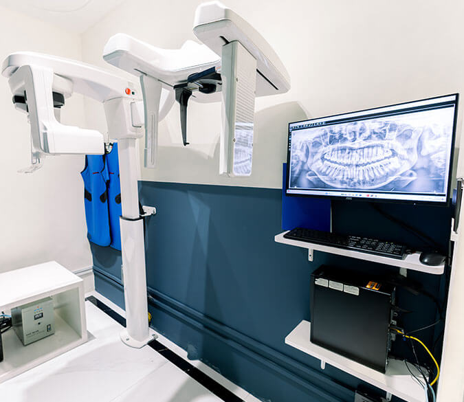
Digital dental x-rays, also known as digital radiography, have revolutionized the field of dentistry by offering a state-of-the-art imaging technology for evaluating a patient’s oral health. Utilizing electronic sensors and low levels of radiation, digital dental x-rays provide computer-generated images of a patient’s internal dental structures. These high-resolution images are essential for identifying and diagnosing oral health issues such as dental cavities, impacted teeth, and other dental abnormalities.
Advantages
-
Instant Imaging and Accessibility
One of the most significant advantages of digital dental x-rays is the speed at which the images can be obtained and viewed. The scans are sent directly to a computer, and dentists can instantly view them on a screen. This not only saves time but also allows for immediate assessment and treatment planning.
-
Elimination of Film Development
With traditional x-rays, film development in a darkroom was required, which could be time-consuming and environmentally unfriendly due to the chemicals used. Digital dental x-rays eliminate the need for film processing, making the process more efficient and environmentally conscious.
-
Enhanced Clarity and Detail
Digital dental x-rays produce clearer and more detailed images compared to traditional x-rays. This improved image quality allows dentists to detect even minor dental issues and provides a more accurate diagnosis.
-
Safer and Lower Radiation Exposure
Digital dental x-rays use lower radiation doses compared to traditional x-rays, making them safer for patients. This is especially important for individuals who require frequent x-rays or for children, who may be more sensitive to radiation.
TYPES
Different Types of Digital Dental X-rays
Panoramic Dental X-ray
A tooth extraction is one of the most common types of oral surgery. This is performed to treat:
- extensive tooth decay that can no longer be saved
- severe mobility caused by gum disease
- dental traumatic injuries
- overcrowding of teeth as a preparation prior to definitive treatments such as braces
Cephalometric Dental X-ray
Periapical Dental X-ray
OUR PROCEDURE
How it Works
01
Examination of the Teeth
The procedure for installing a dental bridge normally takes 2 to 3 separate visits. At the first appointment, the dentist examines the adjacent teeth to make sure that they can support a dental bridge. X-rays of the surrounding tooth and bone are also taken.
02
Preparation of the Teeth
03
Placement of the Temporary Dental Bridge
04
Trial and Installation of the Final Dental Bridge
01
02
03
Once the permanent bridge is ready, the patient is called in for a second visit. During this appointment, the dentist will test the fit, shade, and position of the dental bridge on the patient's mouth. Once everything is perfect, the final bridge is bonded to the teeth using permanent cement. After cementation, the dentist will provide hygiene information to maintain the performance and longevity of the dental bridge and preserve the health of the teeth and gums.
04
FEATURED CASES
Before & After
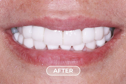
Tooth discoloration is when the color of your teeth change. They don’t look as bright or white as they should. Your teeth may darken, turn from white to different colors, or develop white or dark spots in places.
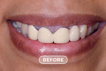
BEFORE
AFTER
Discolored Teeth, Old Restorations
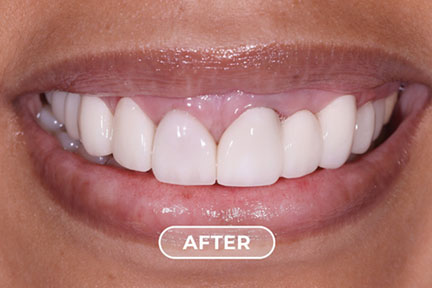
BEFORE
AFTER
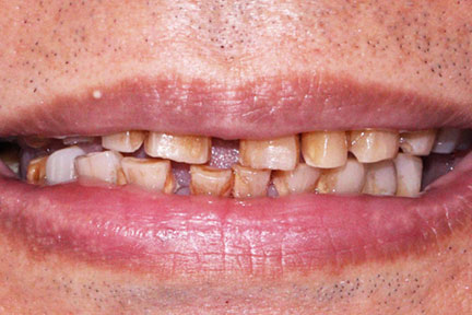
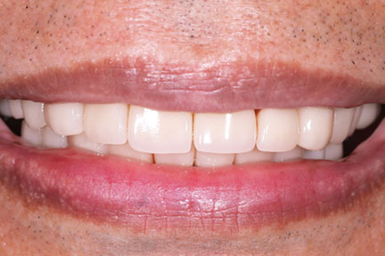
Uneven Teeth Shape, Discolored Teeth
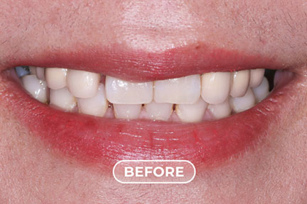
BEFORE
AFTER
Uneven Teeth Shape, Discolored Teeth
ABOUT DENTURES
Frequently Asked Questions
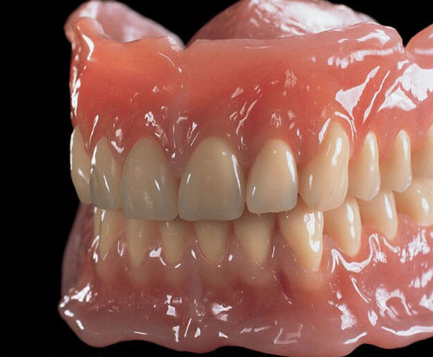
Dentures are custom-made restorations designed to replace missing teeth and restore your smile, bite, and facial support. They are carefully crafted to provide both natural aesthetics and comfortable function.
We offer premium denture materials including Ivocap, Lucitone, Flexite, and ThermoSens, each selected for their durability, comfort, and natural appearance.
- Partial dentures are used when some natural teeth remain, and they are designed to fill the gaps while preserving your existing teeth. Full dentures replace all teeth in the upper or lower arch, restoring complete function and smile aesthetics.
The process typically takes several appointments over a few weeks to ensure precise fit, comfort, and appearance. Each step is customized to achieve optimal function and a natural-looking result.
Yes, dentures allow you to eat and speak more comfortably after tooth loss. There is a short adjustment period, after which most patients regain normal function and confidence.
Dentures typically last 5 to 10 years, depending on the material, fit, and how well they are maintained. Regular dental visits are important to ensure proper fit and long-term comfort.
ABOUT DENTAL IMPLANTS
Care Instructions
Post-Implant Follow-up & Maintenance
After the dental implant procedure, regular check-ups with the dentist every four months are essential. During these follow-up treatments, the dentist will monitor the health of the individual implants, clean around the abutments and dental restorations, and ensure proper oral hygiene is maintained.
Risk Factors & Factors
- Heavy Smoking: Smoking can hinder the healing process and negatively affect the integration of the implant with the jawbone.
- Excessive Alcohol Intake: Alcohol abuse can compromise the body’s ability to heal and may lead to implant failure.
- Periodontal Gum Disease: Untreated gum disease can weaken the surrounding tissues and jeopardize the stability of dental implants.
- Immuno-compromised Individuals: Patients with compromised immune systems may experience difficulties in healing and may have an increased risk of implant failure.
- Teeth Grinding (Bruxism): Habitual teeth grinding or clenching can exert excessive force on the implants, potentially leading to damage or failure.
In conclusion, dental implants offer a revolutionary and effective solution for replacing missing teeth. With their natural appearance, functional benefits, and long-term reliability, dental implants provide patients with the confidence to smile, speak, and eat without worry. By following the proper oral care routine and attending regular dental check-ups, patients can enjoy the full benefits of dental implants for a lifetime.
The Future of Dental Imaging
Digital dental x-rays represent a significant advancement in dental imaging technology, improving diagnostic accuracy and treatment planning. As the technology continues to evolve, it is likely that digital dental x-rays will become even more sophisticated, providing dentists with increasingly detailed and precise images. Additionally, advancements in artificial intelligence and computer-assisted diagnosis may further enhance the efficiency and accuracy of digital dental x-rays, benefiting both dental professionals and patients alike.
In conclusion, digital dental x-rays have transformed the way dentists diagnose and treat oral health conditions. With their speed, clarity, and reduced radiation exposure, digital dental x-rays have become an indispensable tool in modern dentistry.
Not sure which treatment is right for you?